Broken Bones: The Radiologic Atlas of Fractures and Dislocations, 2nd Edition
Felix S. Chew, Catherine Maldijan, Hyojeong Mulcahy
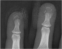
Broken Bones contains 434 individual cases and 1,101 radiologic images illustrating the typical and less typical appearances of fractures and dislocations throughout the body. The first chapter describes fractures and dislocations of the fingers, starting with fractures of the phalangeal tufts and progressing through the distal, middle, and proximal phalanges and the DIP and PIP joints. Subsequent chapters cover the metacarpals, the carpal bones, the radius and ulna, the elbow and upper arm, and the shoulder and thoracic cage. The cervical spine and the thoracic and lumbosacral spine are covered in separate chapters, followed by the pelvis, the femur, the knee and lower leg, the ankle, the tarsal bones, and the metatarsals and toes. The final three chapters cover the face, fractures and dislocations in children, and fractures and dislocations caused by bullets and nonmilitary blasts.
კატეგორია:
წელი:
2016
გამოცემა:
2nd
გამომცემლობა:
Cambridge University Press
ენა:
english
გვერდები:
406
ISBN 10:
1107499232
ISBN 13:
9781107499232
ფაილი:
PDF, 103.62 MB
IPFS:
,
english, 2016
 Amazon
  Barnes & Noble
  Bookshop.org
გსურთ დაამატოთ წიგნის მაღაზია? დაგვიკავშირდით support@z-lib.do-ით
 ფაილების კონვერტაცია
 ძიების მეტი შედეგი
 სხვა უპირატესობები


საკვანძო ფრაზები
დაკავშირებული კოლექციები